Simple Microscope
The Structure of the Simple Microscope
Describe the structure of the simple microscope
A magnifying glass, an ordinary double convex lens with a short focal length, is a simple microscope. The reading lens and hand lens are instruments of this type. When an object is placed nearer such a lens than its principal focus, i.e., within its focal length, an image is produced that is erect and larger than the original object. The image is also virtual; i.e., it cannot be projected on a screen as can a real
image.
image.
The Mode of Action of a Simple Microscope
Describe the mode of action of a simple microscope
The
image formed by magnifying glass or simple microscope is virtual and
erect object place between principal focus (f) and convex lens.
image formed by magnifying glass or simple microscope is virtual and
erect object place between principal focus (f) and convex lens.
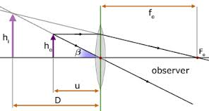
- The normal district vision
- The position of the lens is usually adjusted so that V is about 25cm, which is the shortest distance of distinct vision.
Using the equation of lens (Lens formula).
I/U + I/V = I/F
Adopting the ‘real is positive’ sign convention we obtain:
V = (-Ve) since the image is virtual.
I/U – I/V = I/F
V= 25 –(Normal district vision)
I/U – I/25 =I/F
I/U = I/F + I/25
(I/U)=-1 (25 + F )
25F
U = 25F/F+25
The above formula shows the means of obtaining the distance of object, U.
Magnication (M) of simple microscope
Magnification is the ratio of the image distance to the object distance.
M = Image distance, V
Object distance, U
Hence
M = v/u …………………..(i)
From V = 25cm (distance of district vision)
From U = 25f/(f+25) ……………………… (ii)
Insert eqn (ii) into (i)
M = V/ (25f/(f+25)
M = 25/(25f/f+25)
M = 25/f + 1
Example 1
A
simple microscope with lens of focal length 5cm is used to read
division of a scale 0.5mm in size. How large will the division be seen
through the simple microscope?
simple microscope with lens of focal length 5cm is used to read
division of a scale 0.5mm in size. How large will the division be seen
through the simple microscope?
Data given
- Focal length, f = 5cm
- Required to find magnification, M
Soln:
From
M = (25/f + 1)
= (25/5+1)
=(5+1)
= 6
The magnification of lens = 6
Let the size of the object be ho and that of the image be hi. Then:
M = h1/H ……………(i)
H1 = 6h
The Height , h = (0.5mm)
H1 = 6 (0.5mm)
HI = 3mm
Hence, each division will appear to have a size of 3.0mm viewed through the simple microscope.
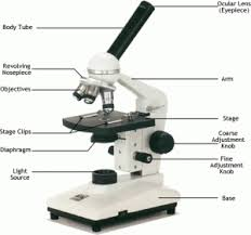
A Simple Microscope
Construct a simple microscope
Parts of simple microscope
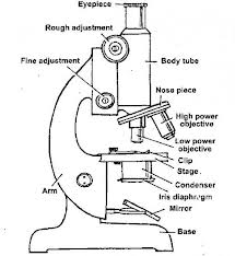
Compound Microscope
The Structure of a Compound Microscope
Describe the structure of a compound microscope
A
compound microscope is an optical instrument used to produce much
greater magnification than that produced by simple microscope. The main
features of a compound microscope includes two short-focus convex
lenses, the objective lens, and the eyepiece.
compound microscope is an optical instrument used to produce much
greater magnification than that produced by simple microscope. The main
features of a compound microscope includes two short-focus convex
lenses, the objective lens, and the eyepiece.
Demonstration
The Mode of Action of a Compound Microscope
Describe the mode of action of a compound microscope
The
most commonly used microscope for general purposes is the standard
compound microscope. It magnifies the size of the object by a complex
system of lens arrangement.
most commonly used microscope for general purposes is the standard
compound microscope. It magnifies the size of the object by a complex
system of lens arrangement.
It
has a series of two lenses; (i) the objective lens close to the object
to be observed and (ii) the ocular lens or eyepiece, through which the
image is viewed by eye. Light from a light source (mirror or electric
lamp) passes through a thin transparent object.
has a series of two lenses; (i) the objective lens close to the object
to be observed and (ii) the ocular lens or eyepiece, through which the
image is viewed by eye. Light from a light source (mirror or electric
lamp) passes through a thin transparent object.
The
objective lens produces a magnified ‘real image’ (first image of the
object). This image is again magnified by the ocular lens (eyepiece) to
obtain a magnified ‘virtual image’ (final image), which can be seen by
eye through the eyepiece. As light passes directly from the source to
the eye through the two lenses, the field of vision is brightly
illuminated. That is why it is a bright-field microscope.
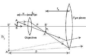
The Magnification of a Compound Microscope
Determine the magnification of a compound microscope
The object lens forms a real and inverted image IIof
the object O ( the image is slightly magnified). The eyepiece lens acts
as a magnifying glass for the first image II and produces a magnifical
virtual image.
the object O ( the image is slightly magnified). The eyepiece lens acts
as a magnifying glass for the first image II and produces a magnifical
virtual image.
The
object is placed just beyond the principal (fo) of the objective lens
so that that the real image I, is formed inside the principal focus (F)
of the eye piece. The eyepiece treats the real image I, as an object and
then forms its magnified virtual image I2.
object is placed just beyond the principal (fo) of the objective lens
so that that the real image I, is formed inside the principal focus (F)
of the eye piece. The eyepiece treats the real image I, as an object and
then forms its magnified virtual image I2.
Magnification of a compound microscope: This
isthe ratio of the image distance produced by a compound microscope to
the object distance. The magnification produced by objective lens is
v/u.
isthe ratio of the image distance produced by a compound microscope to
the object distance. The magnification produced by objective lens is
v/u.
Where
V is the image distance
U is the object distance
The magnification given by the eyepiece is given by;
Me = 25/fe + 1
If the final image is formed at the least distance of distinct vision (V = 25cm).
Mc = Mome
Combine eqn (i) and (ii)
Then
Mc = (v/u) (25/fe+1)
The above formula shows that the final virtual image is formed at the least distance of distinct vision.
Uses of a Compound Microscope
Mention uses of a compound microscope
The uses of a compound microscope includes the following:
- Used to magnify microorganism such as bacteria which cannot be seen by naked eyes.
- Used
in hospitals widely to detect microorganisms in specimens provided by
patients. A specimen is a small amount that is taken for testing. Blood
is an example of specimens. In hospitals microscopes can detect
parasites such as plasmodium ssp (a causative agent for malaria) in
blood specimen.
Example 2
A
certain microscope consists of two converging lenses of focal length
10cm and 4cm for the objective and eyepiece, respectively. The two
lenses are separated by a distance of 30cm. The instrument is focused so
that the final image is at infinity. Calculate the position of the
object and the magnification of the objective lens.
For the objective lens
I/U + I/V = I/Fo
Where
Fo = 10cm
The objective lens forms a real image of the object at the principal focus of the eyepiece.
Thus
V = (30 – 4)
= 26cm
Thus I/U + I/V = I/10
I/U + 1/26 = 1/10
1/U = (1/10 – I/26)
(I/U) -1 = (4/65)
(1/U) -1 = (4/65)-1
U = (65/4)
The distance of object, U= 16.25cm
The magnification given by the objective lense is given by:
Whereas:
V = 26cm
U= 16.25cm
Mo = (26cm/16.25cm)
The magnificent given by objective lens, Mo = 1.6.
Astronomical Telescope
The Structure of an Astronomical Telescope
Describe the structure of an astronomical telescope
An
Astronomical Telescope is used for observing heavenly bodies like stars
and planets (generally bodies which are very far away from normal
vision of human eyes ). Like compound microscope, it consists of two
convex lenses, objective lens and the eyepiece.
Astronomical Telescope is used for observing heavenly bodies like stars
and planets (generally bodies which are very far away from normal
vision of human eyes ). Like compound microscope, it consists of two
convex lenses, objective lens and the eyepiece.
The
focal length Fb of the objective lens is longer than the focal length
Fe of the eye piece lens.Rays of light from a distant object are nearly
parallel when they strike the objective lens of the Telescope.The
objective lens forms a real image, inverted and diminished image IQ of a
distant object is in the focal plane.The eye piece forms the final
magnified image at infinity
When
the telescope is adjusted in such a way that the final image is at
infinity it is said to be in normal adjustment.In this case the distance
between objective lens and eyepiece is (Fb + Fe) This is the maximum
separation between the objective lens and the eyepiece lens.
the telescope is adjusted in such a way that the final image is at
infinity it is said to be in normal adjustment.In this case the distance
between objective lens and eyepiece is (Fb + Fe) This is the maximum
separation between the objective lens and the eyepiece lens.
The Mode of Action of an Astronomical Telescope
Describe the mode of action of an astronomical telescope
The
main reason for a distant object to be smaller is that the two objects
subtend different angles at the eye. In other words, we can say that
different angles substended by the eye causes a distant object to appear
smaller.
main reason for a distant object to be smaller is that the two objects
subtend different angles at the eye. In other words, we can say that
different angles substended by the eye causes a distant object to appear
smaller.
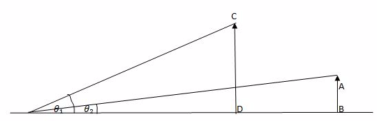
The object AB and CD are of the Same height.The object CD is closer to the eye than AB.
The
object CD appears to be taller than AB because angle B that CD subtends
at the eye is greater than the angle x subtended by AB at the eye.
Images there can be made to appear large by bringing them closer to the
eye.
object CD appears to be taller than AB because angle B that CD subtends
at the eye is greater than the angle x subtended by AB at the eye.
Images there can be made to appear large by bringing them closer to the
eye.
In
a telescope the final image is magnified because it subtends a much
greater angle at the eye than does a distant object observed without a
telescope. B is the angle subtended by the final image at the eye and X
is the angle subtended by a distant object.
a telescope the final image is magnified because it subtends a much
greater angle at the eye than does a distant object observed without a
telescope. B is the angle subtended by the final image at the eye and X
is the angle subtended by a distant object.
The Magnification of an Astronomical Telescope
Determine the magnification of an astronomical telescope
The
magnification of a telescope is defined as the ratio of the angel B (in
radians) subtended by the final image at the eye to the angle X
subtended by a distant object at the eye.
magnification of a telescope is defined as the ratio of the angel B (in
radians) subtended by the final image at the eye to the angle X
subtended by a distant object at the eye.
Thus, for telescope the magnification is given by:
M = B/x ………………………………….i
From figure B= IQ/ID ……………………..ii
X = IQ/IA ………………………………………..iii
But Insert eqn (ii) and (iii) into eqn (i)
M = (12/ID)
(IQ/IA)
M = (IA/ID)
But IA = fo and IF =fe
M = fo/fe……………………………….(x)
Where
Example 3
fois
the focal length of two thin converging lenses of focal lengths 25cm
and 4cm respectively. It is focused on the moon which subtends an angle
of 0.6° at the objective lens. The final image is formed at the
observers least distance of distinct vision (25cm in front of the
eyepiece). Find the diameter of this image.
In the previous figure:
X = h/fo
Where fo is the focal length of the objective lens
X = h/25
Where X is the angle in radians subtended at the objective lens by the moon.
H = 25x
H = 25 (6/10 x 11/180)
H = 25 (6/10 x 22/7 x 1/80)
H = 0.2619m
The height of the image, h = 0.2619m
The distance of this image from the eyepiece is obtained from the relation:
- I/U + I/V = I/fe = 4cm
- V= -25cmV = -25cm
- I/U – I/25 = ¼
- I/U = (1/4 + 1/25)
- (I/U)-1 = (25 +4) -1/100
- 100U= (100/29)
The magnification, m of the lens:
- M = V/u
- M = (25CM/100/29)
- M = 29/4
Let the height of the final image of the moon be h:
- M = Hi/h
- hI = mh
- HI = (29/4) (0.2619)
- HI= 1.90cm
The Height of image Hi = 1.9cm
Hence
The diameter of the final image of the moon will be 1.90cm
Observation
of the universe today are best made from the Hubble Telescope. Outside
the Earth’s atmosphere, this telescope suffer from less interference.
of the universe today are best made from the Hubble Telescope. Outside
the Earth’s atmosphere, this telescope suffer from less interference.
Uses of an Astronomical Telescope
Mention uses of an astronomical telescope
Astronomers use telescopes because they’re much better than our eyes. Here are a few reasons:
- Telescopes
see lots of colours – telescopes can collect light that our eyes are
unable to: radio, microwave, infrared, ultraviolet, x-rays and gamma
rays. - Telescopes collect lots of light – our pupils are only a
few millimeters across, so we can only collect photons over a tiny area
whereas telescopes can collect photons of huge areas (e.g. a football
fields worth for radio telescopes). - Telescopes see fine details
because of the wave nature of light and the nerves in our eyes, we can
only see details about the same angular size as Jupiter’s width.
Telescopes can allow us to resolve fine details – like Jupiter’s Great
Red Spot. - Telescopes can record observations with cameras – You
can see things with your eye and draw them, but telescopes can share
observations with the world! This is especially important for convincing
skeptics that what you saw was real!
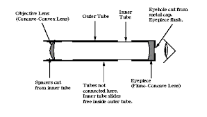
A Simple Astronomical Telescope
Construct a simple astronomical telescope
A simple telescope
Projection Lantern
The Structure of the Projection Lantern
Describe the structure of the projection lantern
The
projection lantern forms images of slides or camera film onto a distant
screen. The film or slide to be projected is inverted and highly
illuminated.
projection lantern forms images of slides or camera film onto a distant
screen. The film or slide to be projected is inverted and highly
illuminated.
The Mode of Action of a Projection Lantern
Describe the mode of action of a projection lantern
Optical arrangement of projection lantern.
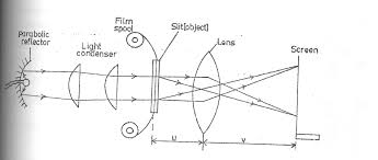
- The slice or film to be projected is inverted and highly illuminated.
- The concave mirror helps to concentrate the light which would otherwise be partly wasted.
- The lamp is placed at the principal focus of the concave mirror.
- The heat filter reduces the heat at falling on the slide or film so as to avoid it overheating.
- Since the image of the projection lantern is Highly magnified, it would not be very bright if there was not enough illumination.
- The
condenser directs a maximum amount of light from the source of the
slide and produce uniform illumination the screen. (The condenser is a
double in order to reduce chromatic aberration). - The projection lens forms the image of the slide on the screen.
- The
light source is usually located at a distance of 2f from a condenser
and invited so that the image on the screen is upright (erect). - The
focal length of the projection lens is ABOUT TWICE THE FOCAL length of
the condenser since the screen is usually far from the lens.
The Magnification of a Projection Lantern
Determine the magnification of a projection lantern
Example 4
A
lantern projector using a slide of (2cm x 2cm) projects a picture (1cm x
1cm) onto a screen 12m from the projection lens. How far from the lens
must the slide be? Find the approximate focal length of the projection
lens.
lantern projector using a slide of (2cm x 2cm) projects a picture (1cm x
1cm) onto a screen 12m from the projection lens. How far from the lens
must the slide be? Find the approximate focal length of the projection
lens.
Solution
The magnification m is given by;
M = V/U ………………………………………i
Where
- HI is the size of image
- H is the size of object
- U object distance
- V image distance
Thus
M = Hi …………………. ii
Then eqn (i) = eqn ii
- v/u =hi/h
- (1200/u)-1 = (100/2) -1
- (u/1200) = (2/100)
- U = (2/100) (1200)
- U = 24cm
The object distance, U = 24cm
Uses of a Projection Lantern
Mention uses of a projection lantern
Projection lantern are used in various areas. These include:
- Projection of films, slides and transparencies,
- projection of opaque objects, i.e. episcopic projection,
- used in searchlights and headlights,
- used in projection apparatus in industry for gauge and screw thread testing,
- used in physical experiments such as projection of a spectrum,
- used in polarisation experiments and interference experiments.
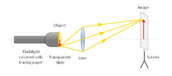
A Simple Projection Lantern
Construct a simple projection lantern
Projection Lantern
The Lens Camera
The Structure of the Lens Camera
Describe the structure of the lens camera
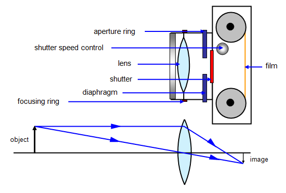
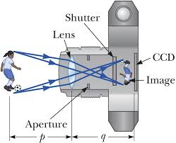
Lens
camera is an instrument which produces an image of object on the screen
using light. The basic physical principle of all camera is the same in
spite of the variation in the design of cameras.
camera is an instrument which produces an image of object on the screen
using light. The basic physical principle of all camera is the same in
spite of the variation in the design of cameras.
The
optical system of the camera are very similar to that of the lantern
projector but with the direction of light reversed.The converging lens
forms a real image of the object to be photographed.(This image is
diminished (smaller than the object and inverted)
optical system of the camera are very similar to that of the lantern
projector but with the direction of light reversed.The converging lens
forms a real image of the object to be photographed.(This image is
diminished (smaller than the object and inverted)
<!–[endif]–>The
lens can be moved back and forward with the help of focusing any so
that objects at different distances can be brought to the focus.A forced
image is locate on the film or plate when the shuttled is open for a
suitable amount of time as determined by the shutter speed.
lens can be moved back and forward with the help of focusing any so
that objects at different distances can be brought to the focus.A forced
image is locate on the film or plate when the shuttled is open for a
suitable amount of time as determined by the shutter speed.
<!–[endif]–>Light enters the camera Box and makes a picture of the object on the film “( The film is sensitive to light)
<!–[endif]–>The
camera is equipped with a diagram or light entering the camera.It
ensures that is incident centrally on the lens so that the distortion of
the image formed is reduced
camera is equipped with a diagram or light entering the camera.It
ensures that is incident centrally on the lens so that the distortion of
the image formed is reduced
The Mode of Action of the Lens Camera
Describe the mode of action of the lens camera
The
aperture stop, which is the limiting diameter of the aperture thought
which light enters the camera (given as fraction of focal length F of
lens) is also called F Number.
aperture stop, which is the limiting diameter of the aperture thought
which light enters the camera (given as fraction of focal length F of
lens) is also called F Number.
This F Number; is the fraction of focal length of the lens given as focal length divide by lens diameter.
F number = Focal length, F/Lens diameter, d
FN = F/d
Where d = is lens diameter.
- The Number Indicates the Number of times the focal length F of times the focal length F of the lens diameter ( or stop)
- The smaller the F – Number for a given focal length the larger the lens diameter
- The lens with a larger diameter has a greater light- gathering power or speed
- This for such a lens the shutter allows light in the camera for a short interval of time.
The Magnification of the Lens Camera
Determine the magnification of the lens camera
Magnification of a lens camerais obtained as the ratio of the Image distance and the object distance.
But from the lens formula:
Thus M = v/U
I/U + I/V = I/F
I/V = I/F – I/U
(I/V) –I = ( U – F / FU)-I
V = FU/ ( U – F)
Example 5
A
lens camera is to be used to take a picture of a man 2m tall if the
lens of the camera Has a focal length of 10cm, calculate the minimum
size of the film frame required, given that the man is 20.1m from the
camera.
lens camera is to be used to take a picture of a man 2m tall if the
lens of the camera Has a focal length of 10cm, calculate the minimum
size of the film frame required, given that the man is 20.1m from the
camera.
Solution:
Magnification is given by:
M = f/ (u-f)
Where
F= 10cm U = 201/m / 2010cm
M = ( 10/2010 – 10)
M = 1/20 ………………………………i
Let the size of the frame be h when the height of man is 2m.
Then
M = 1/200
But h1/h = 1/200
h1 = (1/200) 2
h1 = (1/200)2
h1 (2/200)
h1 = (1/100) m
h1 = 1cm or 10mm
The film frame should be at least 10mm square.
Simple Lens Camera
Construct a simple lens camera
A simple lens camera
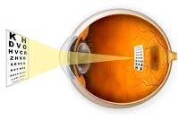
The Structure of the Human Eye
Describe the structure of the human eye
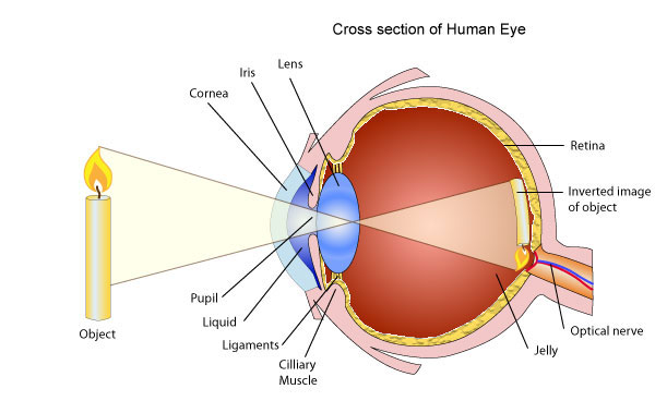
The
eyeball approximately spherical in shape.The wall of this sphere
consist of two layers, the outer layer or sclera and the inner layer or
choroid.The front portion of the SCLERA FORMS A TRANSPARENT CURVED
section called the camera.The choroid layer is balance in order to
prevent internal reflection and also to protect the light sensitive
parts of the eye.
The
aqueous and vitreous hum our are jelly – like substance that fills the
spaces within the eyeball.The aqueous humour is the salt solution of
refractive index n, 1.38.Vitrous hurmour is a watery , Jelly substance
of refractive index 1.34.Behind the cornea there is a colored diagram
called the iris.
aqueous and vitreous hum our are jelly – like substance that fills the
spaces within the eyeball.The aqueous humour is the salt solution of
refractive index n, 1.38.Vitrous hurmour is a watery , Jelly substance
of refractive index 1.34.Behind the cornea there is a colored diagram
called the iris.
The
iris has the central hole called the pupil. The iris contains muscles
which control the size of the pupil. The size of the pupil decreased in
the bright light and increased in the dim light.
iris has the central hole called the pupil. The iris contains muscles
which control the size of the pupil. The size of the pupil decreased in
the bright light and increased in the dim light.
Behind
the pupil and there is a crystalline lens held in position by
suspensory ligaments that are attached to the choroid layer.Near the
suspensory ligaments are the ciliary muscles.The function of the
suspensor ligaments there are the cilliary muscles.
the pupil and there is a crystalline lens held in position by
suspensory ligaments that are attached to the choroid layer.Near the
suspensory ligaments are the ciliary muscles.The function of the
suspensor ligaments there are the cilliary muscles.
The
function of cillary muscles is to control the thickness of the lens.
The lens become thick when the ciliary muscles contract and thin when
the ciliary muscles are relaxed.
function of cillary muscles is to control the thickness of the lens.
The lens become thick when the ciliary muscles contract and thin when
the ciliary muscles are relaxed.
<!–[endif]–>At
the back of the eye there is a retina (This is the part of the eye
which is sensitive to light).Image formed is inverted formed on the
Retina ( This is the part of the eye which is sensitive to light.)
the back of the eye there is a retina (This is the part of the eye
which is sensitive to light).Image formed is inverted formed on the
Retina ( This is the part of the eye which is sensitive to light.)
Image
formed is inverted formed on the retina by successive refraction of
light at the corner, the aqueous hurmour the crystalline lens and the
Vitreous hurmour.Electrical signals are then transmitted to the Brain
through the topic nerve. Finally, the brain interprets these signals.
formed is inverted formed on the retina by successive refraction of
light at the corner, the aqueous hurmour the crystalline lens and the
Vitreous hurmour.Electrical signals are then transmitted to the Brain
through the topic nerve. Finally, the brain interprets these signals.
Accommodation Power of the Human Eye
Explain accommodation power of the human eye
Accommodation is the process whereby the eye alters its focal length in order to form images of objects at different distances.
(Thickening or Thinning of the lens causes a change in its focal length).
The
thickening or thinning of the crystalline lens is made possible by the
action of the ciliary muscles.To view neare object t, ciliuary muscles
contract, this makes the lens thicker.
thickening or thinning of the crystalline lens is made possible by the
action of the ciliary muscles.To view neare object t, ciliuary muscles
contract, this makes the lens thicker.
In
the relaxed state of ciliary muscles, the crystalline lens become
thinner and enables the eye to see (view) distant objects. The farthest
point which can be seen clearly is called the far point of the eye and
the nearest point is called the near point of the eye.
the relaxed state of ciliary muscles, the crystalline lens become
thinner and enables the eye to see (view) distant objects. The farthest
point which can be seen clearly is called the far point of the eye and
the nearest point is called the near point of the eye.
The
corresponding distance from these points to the eye are referred to as
the maximum and least distance of district vision respectively.A normal
eye (i.e. without defects of vision) has a far point at infinity and
near point at a distance of 25cm from the eye.Structure of lens “ view
distant object”
corresponding distance from these points to the eye are referred to as
the maximum and least distance of district vision respectively.A normal
eye (i.e. without defects of vision) has a far point at infinity and
near point at a distance of 25cm from the eye.Structure of lens “ view
distant object”
The Defects of the Human Eye
Identify the defects of the human eye
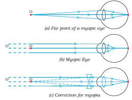
Myopia or near-sightedness
- This defect causes person to see near object clearly while distant objects are not seen clearly.
- The strength of the cornea and the eye lens combination is too great even when muscles of the eye are completely relaxed.
- The focal length of the cornea and the eye – lens combination is always less than the distance to the retina.
- Images
of distant object are formed in front of the retina even when eye is
totally relaxed. However, an object that is closer can be brought into
focus. - In this situation the focal length of the cornea and the
eye lens is so short that objects closer than the conventional (near
point of 25cm) can be brought into focus. That’s why this condition is
called Short sightedness (near sightedness). - Since the problem
is that the strength of the eye – lens and the cornea combination is too
great, the solution is to provide eye glasses (or contract lenses) with
negative lens. - The negative lens weakens the strength of the
cornea and eye – lens just enough so that the resulting focal length
when the eye muscles are relaxed matches the distance back to the retina
so that distant images are now in focused. - The eye glass lenses are negative lenses that means they are thinner in the middle than at the edges.
- It
is easy to identify this kind of eye glass lenses since acting by
themselves they do not form a real image of an object at any distance.
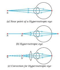
Hyperopia or far-sightedness
- This defect causes a person to see distant objects only and short-distance objects are not seen clearly.
- In
the person with this condition, the strength of the cornea and the
eye-lens combination is too weak when the eye muscles are totally
relaxed. So the image of a distant object is formed behind the retina. - The
solution in the opposite of myopia. Victims should wear positive eye
lenses which strengthen the corner and the eye lens just enough so that
the resulting focal length when the eye is relaxed matches the distance
to the back of the retina.
Astigmatism
- This
occurs when the focal length for the cornea and the eye’s lens for an
object oriented in some direction is not the same as for another located
in a perpendicular direction. - The eye can not bring the
vertical and horizontal lines in a ‘+’ symbol in sharp focus at the same
time. (The axis of differing focal length need not be exactly
horizontal and vertical). - The problem is that the cornea of the
eye lens is not symmetrical. The solution is to use eye glasses whose
lenses are not symmetrical in a complementary way. - The cylindrical lens may be combined with an additional positive or negative lenses.
<!– [if gte mso 9]><xml>Normal
0false
false
false
0false
false
false
EN-US
X-NONE
X-NONE
MicrosoftInternetExplorer4
</xml><![endif]–> Decreased accommodation
- This condition typically occurs in middle-aged people.
- The eye muscles gradually weaken with age, so that the range or accommodation is decreased.
- People with this condition cannot bring both near objects and far objects into focus.
- The
weakening of the eye muscles often causes the focal length of the eye
lens to increase as well so that many people of middle age tend to
become far sighted. - Since the problem is adequate accommodation, no single lens can correct it and people with this problem usual needs bifocals.
- Bifocals are glasses with two different lens strengths, one for near and one for distant objects.
- The usual arrangement is that the bottom half of the lens is the near strength and the top half is the far strength.
The Correction of the Defects of Human Eye
Describe the correction of the defects of human eye
Myopia
is common name for impaired vision in which a person sees near objects
clearly while distant objects appear blurred. In such a defective eye,
the image of a distantobject is formed in front of the retina and not at
the retina itself. Consequently, a nearsighted person cannot focus
clearly on an object farther away thanthe far point for the defective
eye.
This
defect arises because the power of the eye is too great due to the
decrease in focal length of the crystalline lens. This may arise due to
either
defect arises because the power of the eye is too great due to the
decrease in focal length of the crystalline lens. This may arise due to
either
- excessive curvature of the cornea, or
- elongation of the eyeball.
Correction:Thisdefectcan becorrectedby using aconcave (diverging) lens. A concave lens of appropriate power or focal length is able to bring the image of the object back on the retina itself.
Farsightedness,
also called hypermetropia, common name for a defect in vision in which a
person sees near objects with blurred vision, while distant objects
appear in sharp focus. In this case, the image is formed behind the
retina.
also called hypermetropia, common name for a defect in vision in which a
person sees near objects with blurred vision, while distant objects
appear in sharp focus. In this case, the image is formed behind the
retina.
This defect arises because either
- the focal length of the eyelens is too great, or
- the
eyeball becomes too short, so that light rays from the nearby object,
say at point N, cannot be brought to focus on the retina to give a
distinct image.
Correction:This defect can be corrected by using aconvex(converging) lensof
appropriate focal length. When the object is at N’, the eye exerts its
maximum power of accommodation. Eyeglasses with converginglenses supply
the additional focussing power required for forming the image on the
retina.
appropriate focal length. When the object is at N’, the eye exerts its
maximum power of accommodation. Eyeglasses with converginglenses supply
the additional focussing power required for forming the image on the
retina.
The Human Eye and the Lens Camera
Compare the human eye and the lens camera
The camera
- The eye and the camera has a have a convex lens which form a real and inverted image of an object.
- The
eye and the camera are blackened inside to prevent internal reflection.
Rays of light which are not received on the retina or camera film are
absorbed by the choroid layer of the eye or the black surface inside the
camera. - The eye can regulate the amount of light that passes
through the crystalline lens by using pupil while in a camera the
diaphragm regulates light. - In the eye the image is formed in the retina while in the camera the image is formed on the photographic plate.
- The
eye can change the focal length of its lens by the contraction and
relaxation of the ciliary muscles. In this way the eye can focus objects
at different distance. In a camera objects at different distance are
focused on by moving the lens forwards and backwards.