


")


 March 2024 – Primary School Exam")

 MARCH 2024")
, March 2024")
, March 2024")


 March 2024")
 – All Subjects")
 Joining Instruction & Courses Offered | Apply Now")


TOPIC 6: GASEOUS EXCHANGE AND RESPIRATION ~ BIOLOGY FORM 5
Gaseous exchange is the movement of oxygen and carbondioxide between the organism and its environment at the respiratory surfaces.
Oxygen goes in and carbon oxide goes out – These gases move in and out through diffusion. Respiratory surfaces – These are surfaces on which gaseous exchange takes places.
In case of unicellular organisms, gaseous exchange usually takes place throughout the whole body i.e. the cell and the distance through which gases have to travel is small.
In large multicellular organisms there are well developed respiratory systems through which gases move in and out of the organism from and to the external environment.
Types of respiratory surfaces
Skin – amphibians e.g. toads /frogs.
Lungs – chordata and aves.
Lung books– arachnida e.g. scorpion
Gills – external gills e.g. larval amphibians and crustaceous. Internal gills e.g. fishes
Tracheal in insects. Tracheal receive and give out gases directly.
Buccal cavity-amphibians.
Characteristics (Properties) of respiratory surfaces
They should be moist in order to dissolve the gases ( gases diffuse better when they are in solution they must also be permeable)
They must have large surface area to volume ratio to take in or take out gases.
They must be thin to minimize the distance through which the gases have to travel.
They must have a respiratory pigment e.g. Haemoglobin-iron containing pigment; Haemocynine-copper containing pigment.
Ventilation i.e. there must be a constant supply of air or water to the respiratory surface because as oxygen diffuses inwards it tends to be depletal immediately next to the gaseous exchange surface. Therefore the supply of the oxygen from the external environment can be much more efficiently replaced. A flow of this kind is called ventilation.
They must be highly vascularised i.e. supplied with blood capillaries (vessels) to transport the gases.
GASEOUS EXCHANGE IN MAMMALS (MAN)
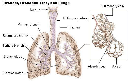
The organ of gaseous exchange is the lung and the respiratory surfaces are the alveoli, the gaseous exchange takes place along the respiratory rate.
Air enters the body through the nose
It then moves to the trachea
The trachea branches into 2 bronchi (bronchus)
The bronchi further branch into several bronchioles. (The bronchioles contain less cartilage than the bronchi).
The respiratory surfaces are the alveoli where gaseous exchange takes place
The two lungs of the body contain about 700million alveoli which can stretch out to about an area of 140m squared(140m2). The two lungs have different size due to the presence of the heart on the left side, to accommodate this area; the lungs have a highly organized internal structure supported by connective tissue. The lungs are compact relatively, firm and highly elastic to allow expansion when filled with air
Features contributing to the efficiency of the respiratory tract.
The walls of the passage/tract are lined with ciliated epithelial cells (have hair like structure) and goblet cells which produce mucus. The mucus traps foreign particles like dust, bacteria which enter the respiratory tract with air. The vibration of the cilia sweeps the trapped particles backwards into the pharynx and then they are swallowed.
Presence of hair in the nasal cavity. This traps the particles coming in along with air. They act as filters of dust and foreign particles.
The mucus produced not only traps the foreign particles but also moistens the respiratory tract.
Presence of numerous blood capillaries whose blood provide a continuous supply of moisture to air to keep it moist before it reaches the alveoli and also supplies heat to warm the incoming air so that the air is warmed up to the body temperature so that the alveoli are not damaged.
The trachea remains open for continuous inhalation and exhalation. This is made possible by the cartilage which also gives strength to the passage.
Presence of collagen and elastic fibres in the alveoli which allow the alveoli to expand and recoil easily.
Kills any bacteria that make it to the alveoli.
It speeds up the transportation of carbon dioxide and oxygen between the air and the liquid lining of alveoli.
It lowers the surface tension of the liquid layer lining the alveoli.
Presence of microphages on the surface of the alveoli which keep them clean by scavenging the bacteria that reaches the alveoli. Microphages are actually what reaches the alveoli. Microphages are actually white blood cells.
INTERNAL STRUCTURE OF THE GASEOUS EXCHANGE PATHWAY TRACT
THE NASAL CAVITY
The nasal passages have a relatively large gaseous area but no gaseous exchange takes place here, the passages have a good blood supply and the lining secretes mucus and it is covered by hair. The air is cleaned moistened and warmed as it passes the nasal cavity.
THE TRACHEA
The trachea is a major airway, heading down to the chest cavity. It is lined with columnar epithelial cells. In the layers below the epithelium are mucus secreting cells (goblet cells). The inner side of the columnar cells is lined with air.
-The trachea is made up of cartilage rings which prevent them from collapsing. The cartilage rings are incomplete to allow the easy passage of food down the oesophagus that runs below the trachea.
– The cartilage rings are C – shapes
THE BRONCHUS
The trachea divides into two bronchi within the chest cavity one leading to the left lung and the other leading to the right.
The bronchi are very similar to the trachea in structure, only that they are narrow The left bronchus divided into two while the right bronchus divided into three.
THE BRONCHIOLES
The bronchioles are much smaller than the bronchi and these are many of them decreasing in size as they go down to the alveoli (different sizes). Larger bronchioles have cartilage rings unlike those which are smaller. These small bronchioles collapse quite easily as the bronchioles get smaller. The lining epithelium changes from columnar to flattened cuboidal cells making diffusion more likely.
THE ALVEOLI
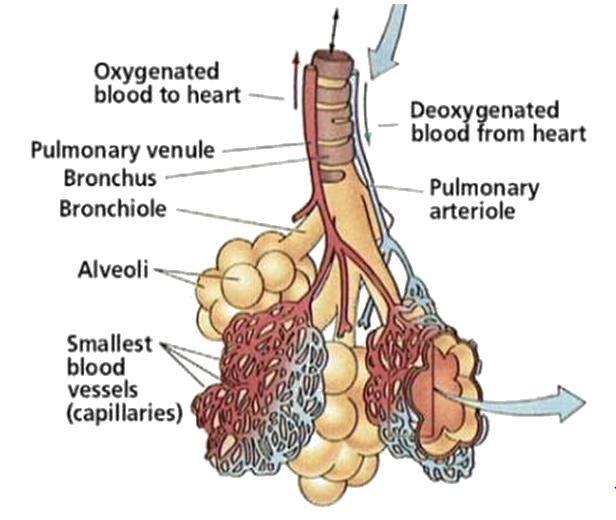
The tiny bronchioles terminate into millions of microscopic air sacs or alveoli in grape like clusters.
The alveoli are 0.1 mm in diameter and they are 0.5 mm in thickness.
They have elastic walls lined (supplied) with blood capillaries from the pulmonary artery
The alveoli are made up of squamous epithelial cells which facilitate diffusion because they have a large surface area and are thin reducing the distance through which gases travel.
The capillaries which run close to the alveoli also have walls which only one cell thick are creating the best possible conditions for gaseous exchange. Between the capillaries and the alveolus is a layer of elastic connective tissues which holds them together.
The elastic elements in the tissue help to force air out of the stretched lungs; this is known as elastic recoil of the lungs.
Breathing in ( inspiration) humans
External intercostal muscles contract and internal intercostal muscles relax.
The ribs move upwards and outwards (anteriorly & ventrally)
The diaphragm muscle contracts and flattens
These two movements of the ribs and diaphragm cause the volume of the thoracic cavity to increase and therefore the pressure inside it falls.
The pressure inside the lungs is lower than that of the atmosphere.
This causes air to rush into the lungs from the exterior.
Breathing out (expiration) in humans
Breathing in is an active process but breathing out is largely passive.
Internal intercostals muscles contract and external intercostals muscles relax
The ribs move downwards (posteriorly) and inwards (dorsally)
The diaphragm muscle relaxes and resumes its dome – shape
These movements cause the volume of the thoracic cavity to decrease and therefore pressure inside increases.
This causes air to be forced out of the lungs as their elastic walls recoil.

TRANSPORTATION OF OXYGEN
There are two ways in which oxygen is transported to the respiring cells. It is carried in the blood in two forms
1. Dissolved oxygen
Oxygen is dissolved in the plasma then transported to the respiring cells. It is a simple physical solution. About 2% of the oxygen in the body is transported in this way.
2. Chemical form
It is transported in combination with haemoglobin. About 98% of the O2 in the body is transported in this way.
THE CHEMICAL FORM
Oxygen is transported in combination with haemoglobin.
Haemoglobin is a protein that consists of four sub- units 2 chains and 2β chains [haemoglobin has four sub – units 2 alpha chains and 2 B chains].
These form in a quaternary structure from the haemoglobin. Each of these four subunits contains a special pigment or Haem group.
The haem group has an iron atom located at its centre. A single Fe atom builds/ binds one molecule of oxygen (02), one molecule of haemoglobin and therefore combines reversibly with up to 4 molecules of oxygen.
Haemoglobin is found in the red blood cells. It has high affinity for oxygen. It combines readily with oxygen.
4. different kinds of oxyhaemoglobin can be formed as follows;

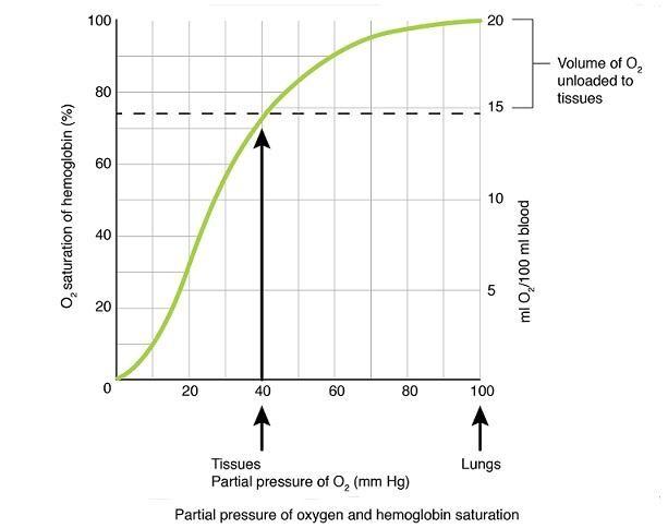
The 4 oxyhaemoglobin differ according to the degree of dissociation Hb4O8 dissociates to release O2 more readily than Hb4O6 and Hb4O6 dissociates more readily than Hb404 and so on.
The degree of dissociation represented gives a character sigmoid (s – shaped) curve called the oxygen dissociation curve.
An oxygen dissociation curve is therefore a curve in which the percentage saturation of haemoglobin is potted against the oxygen concentration (partial pressure)

MYOGLOBIN
Myoglobin is another respiratory pigments in vertebrates
It consists of a single polypeptide chain and a single haem group.
It has an oxygen dissociation curve displaced to the left of the haemoglobin. This means it has higher (greater) affinity for oxygen
THE ADAPTATIONS TO OXYGEN UPTAKE
a) DIVING VERTEBRATES
E.g. Seals, whales, dolphins
The duration of a single dive in seals rarely exceeds 20 minutes where as that of sperm whale may extend to 75 min. Bottle nose dolphin have been known to dive for up to two hours.
The remarkable ability of these mammals to endure such long periods without replenishing the air supplies is a result of
An increased concentration of RBC
Greater haemoglobin concentration
Reduced sensitivity to blood pH
Muscles rich in myoglobin
Reduction in cardiac out put
Restriction of blood supply to vital organs
Tolerance to high lactate levels (muscles respire anaerobically thus accumulation of lactic acid, but they can tolerate lactic acid which would cause fatigue and cramps in other mammals).
Reduction of metabolic rate during diving
Larger tidal volume
Lungs may be entirely collapsed to allow exchange of air (they have few ribs) attached to the sternum making the ribcage more flexible permitting it to collapse partially when under pressures, experienced during a deep dive
Cartileginous rings extend further into the lungs. The rings extend down into the bronchioles to prevent these collapsing under pressures experienced during a deep dive.
Expulsion of air during the dive. This reduces the danger of excessive nitrogen becoming dissolved in the blood (Accumulation of nitrogen, it may dissolve in blood and cause narcotic (harmful nitrogen bubbles)
Closure of the nostrils, anatomical modifications allows the nostrils to be closed during a dive to prevent entry of water into the lungs.
NB:-
As a diver goes deeper, the pressure increases by 1atm for every 10m; in order to pass air from the tanks to lungs its pressure must be increased thus this condition cause greater concentration of oxygen and nitrogen enter the blood.
Oxygen may be toxic and nitrogen has a narcotic effect. Also nitrogen may come out of the solution and form bubbles. If a diver rises to the surface rapidly thus gibe painful symptoms known as bends.
On returning to normal atmospheric pressure the nitrogen dissolves in blood expanding to form bubbles causing pain (bends) and blocking circulation in small blood vessels in the brain and elsewhere.
HIGH ALTITUDE DWELLERS
The amount of oxygen at high altitude levels is the same as that at sea levels. The respiratory problems associated with living at high altitude levels are a result of reduced pressure it means it is more difficult to lead the haemoglobin with O2 effectively.
Some human settlements exist at high altitudes and the inhabitants have become acclimated/ adapted to living in conditions of low atm pressure) the acclimatization involves:-
1. Adjustment of blood pH
The reduced leading of haemoglobin lead to deeper breathing (hyper ventilation in an attempt to compensate for the lack of O2 in the blood). This leads to excessive removal of CO2 and raised blood pH. Nervous responses are triggered causing reduced depth of breathing. In acclimatized individuals the HCO3 ions are removed by the kidney restoring the blood PH to normal (7.4)
2. Increased Oxygen uptake
More O2 is absorbed by the lungs as a result of an improved capillaries network in the lungs and deeper breathing.
3. Improved transport of O2 to the tissues thus results from
Increased RBC conc. (45% – 60% of total blood volume)
The oxygen dissociation curve is shifted to the right to facilitate release of O2 to the tissues
TRANSPORT OF CARBONDIOXIDE (CO2)
The transport of CO2 is closely linked with the transport of O2. CO2 is more soluble in H2O than O2 but its transport in solution is inadequate to meet the needs of most organisms (vertebrates)
CO2 is transported in 3 ways from the tissues (respiring cells) to the gaseous exchange surfaces.
1. Aqueous soln: About 5% of the CO2 is transported in soln in the blood plasma. CO2 is directly dissolved in blood, it occurs in the blood plasma.
1. In combination with haemoglobin: Around 10% of CO2 combines with the amine group in the four polypeptide chains which make up each haemoglobin molecules. It occurs in the RBC
CO2 is transported in the form of carbamino haemoglobin (HbO2)
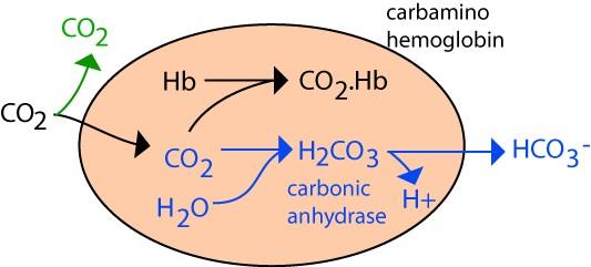

1. In form of hydrogen carbonate (HCO3) 85% of the CO2 produced by tissues combines with water to form carbonic acid

This reaction is catalyzed by a zinc containing enzyme called carbonic anhydrase.
The carbonic acid dissociates to form the hydrogen carbonate ions.

2. The fate of hydrogen ion (H+)
The H+ combines with haemoglobin which loses its O2 . The O2 so released from the Hb diffuses out of the RBC through the capillary wall and tissue fluid into the respiring tissues. Thus the more CO2, the more carbonic acid formed the more H2CO3 dissociates, the more H+ ions released which will combine with the Hb which will then release its O2 to go into the respiring cells. This explains the Bohr’s effect.
Bohr’s effect– the release of oxygen from haemoglobin is facilitated by the presence of carbon dioxide, where CO2 is high in respiring tissues oxygen is released more rapidly.
3. The fate of HCO3–
The HCO3– diffuses out of the RBC into the plasma where they combine with Na+ ions from the dissociation of NaCl to form NaHCO3.
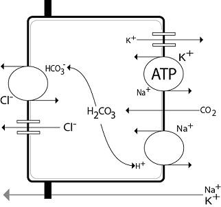
It is largely in this form that the CO2 is carried to gaseous exchange surface; the reverse process is carried out releasing CO2 which diffuses out the body
THE CHLORIDE SHIFT
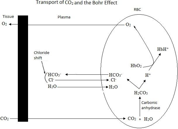
Chloride shift
RESPIRATION
Tissue respiration or cellular respiration or internal respiration is a chemical processes in which food substances such as carbohydrates, lipids and proteins are oxidized in the cells to yield energy.
During respiration the energy that was fixed into the synthesized organic matter is released and made available for use the living organisms.
The Site of Respiration:
The site of respiration depends on the step of the process involved:
lower-roman;margin-left: 17.250000000000007pt;padding-left: 23.15pt;text-decoration: none;vertical-align: baseline”>
lower-roman;margin-left: 17.250000000000007pt;padding-left: 23.15pt;text-decoration: none;vertical-align: baseline”>
Respiratory Substrate:
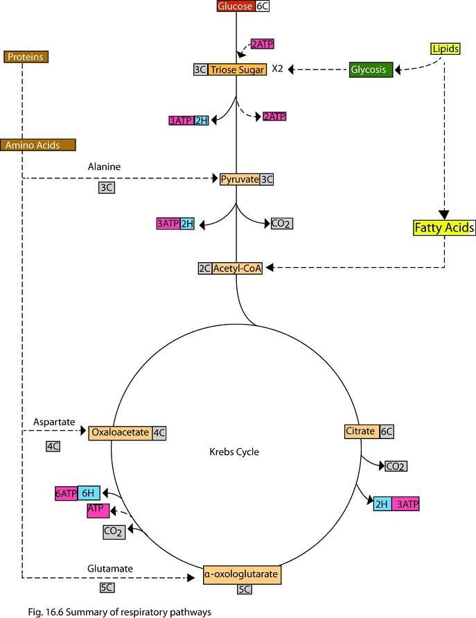
There are food substances which upon being oxidized yield energy in form of ATP, they include carbohydrates, lipids and proteins. However the most preferred respiratory substrates are the carbohydrate especially glucose.
Respiration of Glucose:
Glucose is the mostly used respiratory substances. When glues completely oxidized aerobically a molecule of glucose yield 38 ATP molecules.
STAGES OF RESPIRATION:-
Respiration is completed in the following stages:
lower-roman;margin-left: 17.250000000000007pt;padding-left: 23.15pt;text-decoration: none;vertical-align: baseline”>
lower-roman;margin-left: 17.250000000000007pt;padding-left: 23.15pt;text-decoration: none;vertical-align: baseline”>
lower-roman;margin-left: 17.250000000000007pt;padding-left: 23.15pt;text-decoration: none;vertical-align: baseline”>
lower-roman;margin-left: 17.250000000000007pt;padding-left: 23.15pt;text-decoration: none;vertical-align: baseline”>
GLYCOLYSIS:
Glyolysis (glycol – sugar lyses broken down)
Is the breakdown of hexose sugar, usually glucose into two molecule of 3 – carbon compound pyruwate (pyruwic acid). It sews in a all cell in anaerobic organisms it is the only stage of respiration.
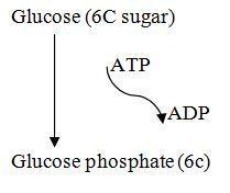
Initially the glucose is insufficiently reactive and so it is phosphotylated poor to split into two trios sugar molecules.
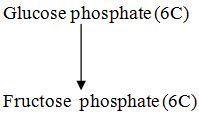

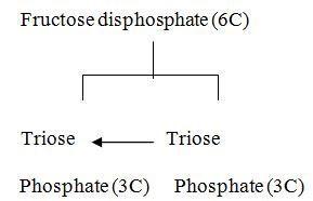
Inorganic phosphate is added to further activate the triose phosphate.


The energy yield is a net gain of two molecules of ATP.
The total yield of energy therefore two molecule of ATP directly and six molecule of ATP produced from the two reduced NAD molecules. A total of eight ATP molecule.
KREBS (tricarboxylic acid) CYCLE:
The pyruvic acid formed as a result of glycolysis may in the absence of oxygen being converted to a variety of substances to yield a little energy. This is anaerobic pathways.
In the presence of oxygen the pyruvic acid enters the Krebs cycle.
Before entering the actual cycle one of the 3-C atom of pyruvic acid is oxidized to CO2 and a molecule of NA is reduced by addition of two hydrogen atom.
This leaves the acetyl group (CH3 CO) which is readily accepted by a coenzyme called coenzyme A.
The two carbon acetyl group of this compound combine with 4 – carbon substance called oxaloacetic acid to give a six carbon molecule atric acid.
In a series of reaction two carbondioxide produced and 4 – carbon oxaloacetic acid regenerated in readness to receive another 2 – carbon acetyl group from acetyl coenzyme A. other products includes a total of 8 hydrogen atoms which are used to reduce three molecules of NAD and one molecule of FAD.
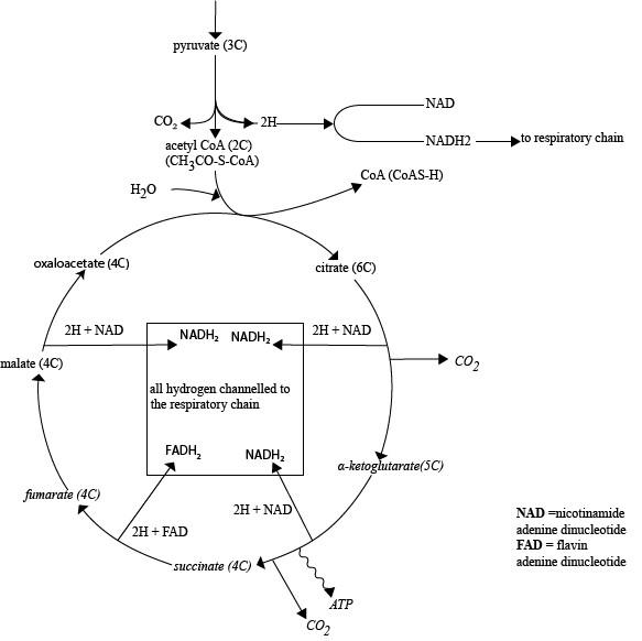
These reduced electron carriers (NAD & FAD) eventually pass on the hydrogen atoms to oxygen yielding 11 more ATP molecule for each pyruvic acid. In addition of further ATP molecule is yielded. Directly during the cycle to give a total of 12 ATB per pyruvic acid molecule.
Importance of Krebs’s cycle:
It provide hydrogen atoms which ultimately yield the major part of the energy derived from the oxidation of glucose molecule.
It is a valuable source of intermediate which are used to manufacture other substances pg. Fatty acids, amino acids, carotenoids.
ELECTRON TRANSPORT SYSTEM:
Is the means by which the energy From the Krebs cycle inform of hydrogen atom is converted to ATP.
Much of the energy is in the form of hydrogen atoms which are attached is the hydrogen carries NAD and FAD.
These atoms passed along a serves of carriers at progressively cover energy levels, as they lose their energy it is harnessed to produce ATP molecule.
There for each molecule of NAD and two for each one FAD
The other camers in the system are iron containing protein called cytochromes. The hydrogen split into their protons and electrons during the pathway.
They combine with their proton before the find stage where the newly reformed hydrogen atoms combine with oxygen to form water.
It is very essential in aviabic respiration only play a role at this final stage, it is vital since it drives the process.

Summary of the Electron transport system.
In absence of oxygen only anaerobic resp. continues the transfer of hydrogen atom is catalyzed by the enzyme cytochrome oxidize. This enzyme is intrubited by cyanide, so preventing the removal of hydrogen atoms at the end of the respiratory chain. In these circumstances the hydrogen atoms accumulate and aerobic respiratory ceases, making cyanide a most effective respiratory incubator.
Anaerobic Pathways:-
If no oxygen is available the pyruvic acid formed at the glycolysis do not enter the Krebs cycle but follow one of the anaerobic pathways are often reffered as fermentation.
Three major anaerobic pathways:-

Yeast and other plants. This process forms the basis of brewing and baking
C. Lactic acid fermentation
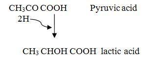
This happen in higher animals especially in middles when oxygen used exceeds supply.
The role of vitamin B complex in cellular respiration
B1 – Thiamine : – Involve in formation of some Krebs’s enzymes
Forms a part of acetyl coenzyme A.
B2 – Riboflavin: – Form part of hydrogen carrier flavo protein (FP)
B3 – Niacic: – Forms part of coenzymes NAD & NADP
(Nuotinic acid) Form part of acetyl coenzyme A
B5 – Partothenic acid
Form part of acetyl coenzyme A
TO COMPLETE TOTAL YIELD OF ATP WHEN OXYGEN RESPIRE
AEROBICALLY
ATP and energy yields
ATP is the form in which energy from the breakdown of glucose is temporarily stored.
When glucose is completely oxidized aerobically the sources of ATP are:-
lower-roman;margin-left: 54pt;padding-left: 23.299999999999997pt;text-decoration: none;vertical-align: baseline”>
lower-roman;margin-left: 54pt;padding-left: 23.299999999999997pt;text-decoration: none;vertical-align: baseline”>
lower-roman;margin-left: 54pt;padding-left: 23.299999999999997pt;text-decoration: none;vertical-align: baseline”>
molecules NADH2 and FADH2
lower-alpha;margin-left: 54pt;padding-left: 2.4000000000000057pt;text-decoration: none;vertical-align: baseline”>
lower-alpha;margin-left: 54pt;padding-left: 2.4000000000000057pt;text-decoration: none;vertical-align: baseline”>
lower-alpha;margin-left: 54pt;padding-left: 2.4000000000000057pt;text-decoration: none;vertical-align: baseline”>
Therefore there are total of 10NADH2 and 2FADH2 molecules.
Each pair of atoms carried by NAD produces in respiratory chain as NADH2, shunts its hydrogen to carrier 1. Where NAD occurs and each pair of hydrogen atom carried by FAD shunts into hydrogen to carrier 2 where FAD occurs.
Thus 10NADH2 produce a total of 3ATP X 10NADH2
= 30 ATP
2FADH2 produce a total of 2FADH2 × 2ATP
= 4 ATP
Summation
From Glycolysis = 2 ATP
From Krebs’s cycle = 2 ATP
From Respiratory chain 34 ATP = 34 ATP
38 ATPS
Thus when a molecule of glucoseis completely oxideized aerobically a total of 38 ATP molecules is synthesized.
The total ATP produced in one
stage: ATP produced directly ATP indirectly Total
Glycolysis 2ATPs 6ATPs
Krebs cycle 2 ATPs 22 ATPs
Pyruvate to acetate – 6 ATPs
Total 4 ATPs 34 ATPs 38 ATPs
TOTAL YIELD OF ATP WHEN GLUCOSE RESPIRES ANAEROBIC RESPIRATION
We have seen in the previous section that only glycolysis occurs during anaerobiosis and that the NADH + H+ it yields is not available for oxidative phosphorylation.
The total energy released is therefore restricted to the two ATP’s formed directly.
In lactate fermentation all is not lost, and the lactate may be converted to pyruvate by the liver and so enter Krebs cycle thus releasing the remaining energy.
RESPIRATORY PATHWAY USING LIPID AND PROTEIN
Sugar are not only material which can be oxidized by cells to release energy. Both fats and protein in certain circumstances be used as a respiratory substrates.
Respiration of Fats:
Fats are used as respiratory substrate when there is unsufficient amount of carbohydrates the oxidation of fats is proceeded by its hydrolysis to glycerol and fat acids.
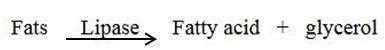
GLYCEROL:
Glycerol is phosphatylated by an isnorganic phosphate to form glycerate phosphate (GP). Then oxidized by NAD to form dehydroxy acetone which is then converted its isomer 3PGAL then feel into the glycolytic pathway at the point where PGAL occurs.
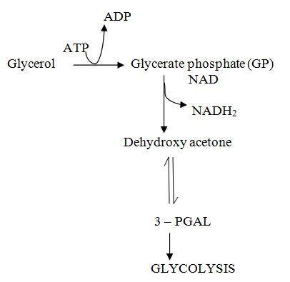
The NADH2 then passed to the respiratory chain with a total yield of 3ATP molecules. The PGAL is passed to the glycolytic and Krebs cycle pathway with a total yield of 17ATP molecules. Thus the net gain of ATP when glycerol is aerobically oxidized is 20 – 3 ATP.
FATTY ACIDS:
Each fatty acid in the matrix of mitochondria undergoes oxidation in the process called βoxidation involves the fragmentation of fatty acid to 2-cabon fragments. Each of these will be converted into acetyl Co-A and then fed into the Krebs at the point where acetyl Co-A occurs.
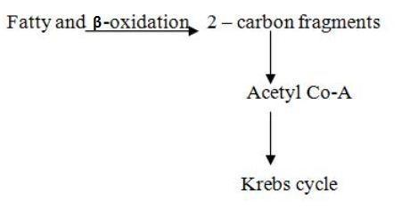
The advantage of respiring fats is that fatty acid have large number of hydrogen atom which when passed through respiratory chain yield a large amount of ATP molecule.
E.g. The respiration of asteric acid, fatty acid and animal adipose tissue yields a total of 147 ATP molecules, Total number of ATP’s formed are 166 ATP’s from glycezid and fatty acids.
Respiration of Protein:
Protein is respired only when both carbohydrates fats are totally absent or need up is used in the condition of starvation. When proteins are to the respired first hydrolysed to amino acids, then diamination.
Oxidative Diamination:
This process occurs in the inner cells and it involves the removal of ammonia from amino acid. This is by dehydrogenation and hydrolysis.
Consider the diamination of glutaric acid
Glutaric acid + NAD + H2O α-Ketiglutaric acid + NADH2 + NH3
The ammonia is then exceted as either wric acid urea or pure NH3 depend on the nature of the environment the diaminated amino acid is converted into one the Krebs cycle intermediate depending on the number of carbon atoms. If it is a 5-carbon amino acid. It will be converted into α- ketoglutaric acid.
And if it is a 4-carbon amino acid will be converted into oxaloacetic acid. If it is a 3-carbon amino acid will be converted into pyruvic acid, latter converted into acetyl COA then fed into Krebs cycle.
Transamination:
This is a process whereby on amino group from one amino acid is transferred to a keto group of another amino acid so as to form a new amino acid. Hence the conversion of one amino acid into another is controlled by transaminase oenym and it can produce α-keto acids that directly enter Kreb’s cycle.
Study questions:
RESPIRATORY QUOTIENT (RQ)
Respiratory quotient is a ratio of the volume of Carbondioxide evolved to oxygen consumed over a period of time.
RQ = Volume of CO2 evolved
Volume of O2 consumed
Significance of RQ:
The value of RQ tells the type of a substrate that is being oxidized
Example (a) For glucose the value f RQ = 1
C6H12 O6 + 602 6CO2 + 6H2O
RQ = Vol of CO2
Vol of O2
= 6 CO2
6 O2
= 1
lower-alpha;margin-left: 27pt;text-decoration: none;vertical-align: baseline”>
Consider the respiration of asteric acid.
C18H36O2 + 2602 18C02 + 18 H20
RQ = Vol of C02 Vol of 02
= 18 C02
26 02
= 0.7
lower-alpha;margin-left: 27pt;text-decoration: none;vertical-align: baseline”>
However in practice the above theoretically, call caulated values cannot be obtain this is because:-
lower-roman;margin-left: 49.60000000000001pt;padding-left: 23.200000000000003pt;text-decoration: none;vertical-align: baseline”>
lower-roman;margin-left: 49.60000000000001pt;padding-left: 23.200000000000003pt;text-decoration: none;vertical-align: baseline”>
2. The value of RQ tells the type of metabolism which is taking place.
lower-roman;margin-left: 36.00000000000001pt;padding-left: 22.9pt;text-decoration: none;vertical-align: baseline”>
lower-roman;margin-left: 36.00000000000001pt;padding-left: 22.9pt;text-decoration: none;vertical-align: baseline”>
lower-roman;margin-left: 36.00000000000001pt;padding-left: 22.9pt;text-decoration: none;vertical-align: baseline”>
lower-alpha;margin-left: 36pt;padding-left: 2.4000000000000057pt;text-decoration: none;vertical-align: baseline”>
lower-alpha;margin-left: 36pt;padding-left: 2.4000000000000057pt;text-decoration: none;vertical-align: baseline”>
lower-alpha;margin-left: 36pt;padding-left: 2.4000000000000057pt;text-decoration: none;vertical-align: baseline”>
Qn: what is the RQ of a photosynthesis green plant at its compensation points?
What will be the nature of the RQ values when aerobic and anaerobic respiration are occurring together
BASAL METABOLIC RATE (BMR)
BMR is defined as the minimum rate of energy conversion required just to stay alive during absolute rest or sleep.
Because it is difficult to ensure a subject is absolute rate the BUR is usually estimated as the amount of energy used by a pelscon restring quietly after at least 13 hours or sleep and 12 hours after the last meal.
The BMR does not remain constant through out life but changes as growth development and aging take place e.g. Newly BMR is 220 and 220 KJM-1 hr. By the end of the year one.
The BMR also varies with sex, and health of the individual for a healthy young women. It is about 150 KJMol-2 hr-1 and for the health young man it is about 170 KJ mol-2 hr-1


 TIMETABLE, MAY 2024")


[url=http://cheapdrugs.store/#]treatment for ed[/url]